What is Gram Stain Test – Staining Procedure, Principle and Results
Last reviewed by Editorial Team on February 9th, 2019.
What is Gram Staining
Gram staining is a microbiologic procedure used to differentiate Gram-negative from Gram-positive bacteria.
It was developed by Hans Christian Gram; a Danish physician, in 1884. The cells are colored red or violet so as to distinguish its group. A microorganism is gram-positive if the stain remains violet secondary to the presence of peptidoglycan in the cell wall.
On the other hand, a microorganism is gram-negative if the stain is red secondary to the thin peptidoglycan wall. (1, 2)
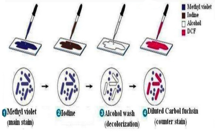
Picture 1 : The image shows the steps needed to complete the gram staining procedure.
Picture Source: vignette.wikia.nocookie.net
Gram staining procedure
There are three processes involved in gram staining. They are the following:
- Staining using a water-soluble dye (crystal violet)
- Decolorization
- Counterstaining using safranin (3)
What you will need?
- Crystal violet which acts as the primary stain
- Gram’s iodine
- Decolorizer in the form of acetone or ethanol
- Secondary stain/counterstain in the form of safranin (4)
- water
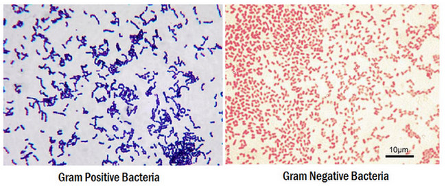
Picture 2: A gram stain procedure showed gram positive bacteria on the left and gram negative bacteria on the right.
(gram stain procedure gran negative positive bacteria)
Photo Source: static1.squarespace.com
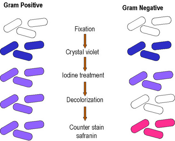
Photo 3: Gram positive bacteria remain purple in color while gram negative bacteria turn pink.
(how does gram positive and gram negative looks like)
Image Source: 2.bp.blogspot.com
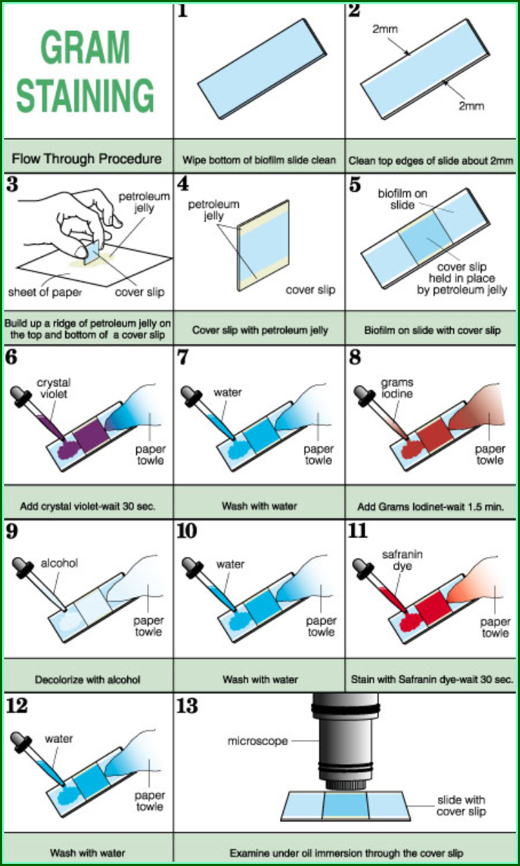
Steps:
- Prepare a slide of a cell sample. Put a drop of sample on the slide and pass it through a Bunsen burner three times. The microorganism to be tested is stained with a crystal violet dye and incubate for a minute.
- Rinse the slide with water for five seconds to get rid of unbound crystal violet dye.
- An iodine solution (a combination of iodine and potassium iodide) is added to form a complex (a large molecule that is insoluble in water).
- A decolorizer like acetone or ethyl alcohol is added and rinse with a gentle stream of water. The purpose of decolorizer is to dehydrate the peptidoglycan layer of the organism. Eventually, the layer shrinks and tightens.
- A gram-positive microorganism is resistant to the large crystal violet iodine complex. The substance cannot penetrate to the peptidoglycan layer.
- A gram-negative microorganism has a thin peptidoglycan layer and so the large crystal violet iodine complex can easily penetrate to it resulting to the lost in color.
- A water-soluble safranin is added to the sample for counterstaining. The secondary stain should be on the slide for a minute and then rinse with a gentle stream of water for about five seconds. It causes the sample to change its color into red. A gram-negative microorganism turns red but the gram-positive bacteria remain violet/purple. (3, 4, 5, 6)
Picture 4 : Gram staining and the steps needed to complete the procedure.
(Gram staining and the steps)
Picture Source: www.cs.montana.edu
Gram staining principle
The composition of the cellular wall of gram-positive and gram-negative bacteria tells the gram staining differences. The following principles apply:
- Bacteria that are gram-positive have a thick layer of peptidoglycan.
- They also have several teichoic acids which are resistant to decolorization.
- The crystal violet solution dissociates into Cl and CV+ and deeply penetrate through the wall and membrane of the microorganisms being tested.
- CV+ interacts with the negatively charged part of the microorganism’s cell leading to a purple stain. Iodine is added and interacts with the CV+ forming a large crystal violet-iodine complex within the cell’s cytoplasm and outer layers.
- A decolorizing agent is added and will interact with the lipids of the cellular membrane of the microorganism being tested.
- The peptidoglycan layer of the gram-negative bacteria is exposed because they have thin peptidoglycan layers.
- As they are exposed to ethanol, the cell wall of gram-negative bacteria leaks thereby allowing the large CV-I complexes to be washed from the cell.
- Gram-positive bacteria have highly cross-linked and multi-layered peptidoglycan which become dehydrated when ethanol is added. As a result, the large CV-I complexes within the cells are trapped leading to purple color when decolorized.
- During decolorization, gram-negative bacteria lose its purple color while gram-positive bacteria retain its color. The difference in color can only be seen during counterstaining. (3, 5, 6, 7)
Why is gram staining important?
The gram-staining procedure is one of the most important procedures in microbiology. Its primary purpose is to differentiate gram-positive from gram-negative bacteria.
Identifying and classifying bacteria are important as they help in the presumptive diagnosis of various types of diseases.
It also helps in understanding the chemical makeup of the cell wall of bacteria which is helpful in determining the course of treatment for a specific type of disease. (3, 8, 9)
Reporting gram staining results
- A gram reaction of bacteria (gram-negative or gram-positive)
- Number of bacteria (scant, few, moderate, or many)
- Morphology of bacteria
- Presence and number of pus cells, yeast cells, and epithelial cells
A gram stain procedure is ordered by the doctor if he/she suspects of infection.
The result of the test can help the doctor in identifying the type of bacteria that cause the infection and determining the appropriate treatment plan. (1, 6, 9, 10)
References:
- https://serc.carleton.edu/microbelife/research_methods/microscopy/gramstain.html
- https://en.wikipedia.org/wiki/Gram_stain
- https://microbeonline.com/gram-staining-principle-procedure-results/
- https://labtestsonline.org/tests/gram-stain
- http://www.austincc.edu/microbugz/gram_stain.php
- https://www.microscope.com/education-center/how-to-guides/grams-stain/
- https://microbiologyinfo.com/gram-staining-principle-procedure-interpretation-examples-and-animation/
- http://www.uphs.upenn.edu/bugdrug/antibiotic_manual/gram1.htm
- https://www.jove.com/science-education/10092/gram-staining-of-bacteria-from-environmental-sources
- https://www.verywellhealth.com/information-about-gram-stain-1958832
