PAS Stain (Periodic acid–Schiff Stain)- Procedure and Uses
Last reviewed by Editorial Team on February 27th, 2019.
What is a PAS Stain?
A PAS Stain is a staining method that detects polysaccharides and mucosubstances.
Examples are glycogen, glycolipids, glycoproteins, and mucins. It stands for Periodic Acid-Schiff stain.
It is one of the commonly used procedures in the histopathology laboratory as it can highlight molecules with high carbohydrate content.
These include glycogen, fungi, mucin, as well as the basement membrane in the skin. (1, 2, 3)
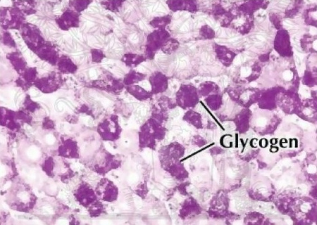
Image 1: A positive polysaccharide result as shown in the discoloration; characterized by magenta color.
Picture Source: laboratoryinfo.com
What is the PAS stain used for?
- It stains structures that contain a high level of macromolecules of carbohydrates which are usually present in connective tissues, basal laminae, mucus, and glycocalyx.
- It is helpful in diagnosing various medical conditions like
- Paget’s disease of the breast
- Glycogen storage disease
- Adenocarcinoma
- Staining macrophages in Whipple’s disease
- Fungal infection
- Alveolar soft part sarcoma
- Erythroleukemia (cancer of the blood secondary to immature red blood cells)
- α1-antitrypsin deficiency
- Ewing sarcoma
- Pulmonary alveolar proteinosis
- Identification of glycogen in biopsy of the lungs in pediatric patients with pulmonary interstitial glycogenosis
- Ceroid lipofuscinosis (it highlights super cross-linked lipids inclusions) (3, 4, 5, 6)
How it is performed?
Ingredients/Materials needed
Periodic acid solution
- Periodic acid
- Distilled water
Schiff’s Reagent
- Fuchsin basic
- Sodium metabisulphite
- Distilled water
- Activated charcoal
- Conc. HCl (5, 6)
Procedure
- Bring the sections to the distilled water and treat with a periodic acid for about five minutes.
- Rinse with distilled water.
- Cover it with Schiff’s reagent for about 15 minutes
- Rinse in running tap water for about 10 minutes.
- Counterstain using Herri’s hematoxylin for about 15 seconds.
- If needed, differentiate using acid alcohol and bluing.
- Rinse in tap water and with an increasing concentration of alcohol.
- Clear in xylene and mount. (6, 7, 8, 9)
PAS Stain Principle
- Initial reaction – The first reaction makes use of a periodic acid that acts as an oxidizing agent. It oxidizes the carbon to carbon bonds between two adjacent hydroxyl groups. The end result is the Schiff reactive aldehyde groups.
- Second reaction – A Schiff reagent will be used and the tissue section will react to the reagent. The reagent is a mixture of sodium metabisulphite, hydrochloric acid, and basic fuchsin. The basic fuchsin present in the mixture will react to the aldehyde groups in the tissues. You will notice a reaction when a bright magenta color is formed.
- Final reaction – If the section is rinsed in water, the fuchsin molecule will produce a bright magenta color. The brighter the color the more hydroxyl groups are there. In some instances, other tissue elements present in the section are visualized using a counterstain agent; the haematoxylin. A light green counterstain is typically used to demonstrate fungal organisms. (4, 7, 9, 10)

Picture 2: A PAS staining procedure showing numerous fungus.
Photo Source: www.researchgate.net

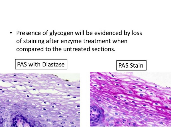
Photo 3: Two PAS Stain procedure; one containing enzymes and the other one without enzymes.
Image Source: www.newcomersupply.com
Interpreting the results
PAS Stain color and the corresponding histologic features:
- Magenta – basement membranes, glycogen, fungal organisms, and mucus substances.
- Blue – nuclei
- Green – other tissue elements (2, 5, 6)
Modifications

Image 4: A PAS stain procedure with diastase.
Picture Source: upload.wikimedia.org
Picture 5: A PAS stain procedure; one is plain staining and the other is with diastase.
Photo Source: image.slidesharecdn.com
A PAS Stain can be done with diastase. Its primary purpose is to differentiate glycogen from other PAS positive elements that can be present in the tissue sample. A perfect example is a mucin.
It can only be identified if glycogen is digested and washed out with diastase.
Such kind of procedure can also be used in differentiating glycogen granules from other granules such as in the case of people with a tumor. (3, 7, 9, 10)
References:
- https://en.wikipedia.org/wiki/Periodic_acid%E2%80%93Schiff_stain
- http://www.pathologyoutlines.com/topic/stainspas.html
- https://bitesizebio.com/13413/why-pick-pas-for-histology/
- https://www.abcam.com/periodic-acid-schiff-pas-stain-kit-mucin-stain-ab150680.html
- https://laboratoryinfo.com/periodic-acid-schiff-pas-staining-technique-for-carbohydrates/
- http://pages.jh.edu/~iic/resources/ewExternalFiles/Glycogen_Staining.pdf
- https://www.sciencedirect.com/topics/medicine-and-dentistry/periodic-acid-schiff-stain
- https://www.sigmaaldrich.com/content/dam/sigma-aldrich/docs/Sigma/General_Information/2/395.pdf
- https://www.nature.com/articles/1971306a0
- https://www.agilent.com/en/product/special-stains/special-stains-kits/periodic-acid-schiff-(pas)-stain-kit-artisan-76940
Similar Posts:
- What is Gram Stain Test – Staining Procedure, Principle and Results
- Benedict’s Test
- Methyl Red Test
- Oxidase Test
- Indole Test
