Buffy Coat
Last reviewed by Editorial Team on February 9th, 2019.
Blood contains millions of cells and they are suspended in the plasma (the liquid part of the blood).
The components of blood include:
- Red blood cells
- White blood cells
- Platelets
The red blood cells contain the nutrients. White blood cells or also known as leukocytes are a significant part of the immune system.
They protect the body from infection and other foreign substances. Leukocyte concentrates are available from the blood.
Platelets serve as the clotting agent. It is easy to examine red blood cells as there are many of them. In a single drop of blood, there are thousands of red blood cells.
On the other hand, white blood cells are of small quantity, which makes examination quite difficult. (1, 4, and 6)
How to check for white blood cell components?
To check for the components of white blood cells, the fastest possible way is to check for buffy coat smear. It is simply the concentration of white blood cells including platelet in the blood sample.
For small volume of blood, leukocytes can be separated through density centrifugation.
What is Buffy coat ?
The procedure is leukocyte concentrate or popularly known as buffy coat. It is called the buffy coat because of its buff hue. The buffy coat pertains to the fraction of the anti-coagulated blood sample.
It contains white blood cells and platelets that produced from density gradient centrifugation of the blood.
To check for a buffy coat, a special machine is used to spin the blood sample in a small circle (centrifugation process). The force causes the blood to separate into three parts.
- Red blood cells settle at the bottom.
- The top layer contains the plasma.
- The white blood cells and platelets will be in the middle, which forms a narrow band. It has a distinct creamy white to buff color, which refers to as the buffy coat. The buffy coat is collected and spread on a clean glass slide. Its components will be checked under the microscope. (1, 2, 3, 4, and 5)
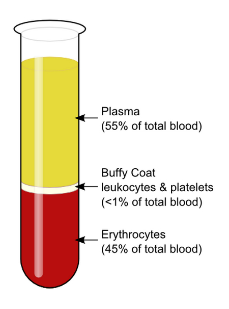
Picture 1 : A blood sample in a test tube. The bottom layer is the red blood cell. The top layer is the plasma and the one in the middle is the buffy coat, which is a combination of leukocytes and platelets.
Picture source : upload.wikimedia.org
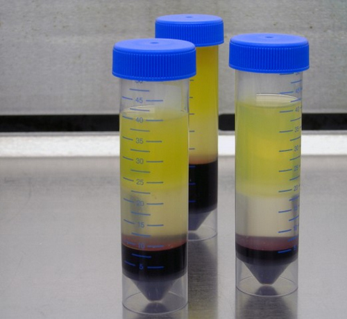
Picture 2 : The samples contain fresh human blood buffy coat.
Picture source : bioscience.co.uk

Picture 3 : Three specimens; one in a healthy state, another one in reactive state, and the last specimen is positive of leukemia.
Picture source : medsci.indiana.edu
What is the diagnostic significance of buffy coat?
- It is useful in extracting DNA from the blood of mammals.
- Quantitative buffy coat helps detect infection with malaria and other blood parasites.
- A buffy coat can be used in many stem cell technologies (usually a part of stem cell technology cell separation kits).
- It helps detect immature or abnormal white blood cells circulating in the bloodstream.
- It is used for the isolation of cells.
- It helps prevent blood transfusion complications. (7)
- It determines cell count in the whole blood.
- A quantitative buffy coat is helpful in detecting malarial parasite in peripheral blood.
- It is a useful tool in diagnosing filariasis.
- A quantitative buffy coat is helpful in diagnosing difficult cases of visceral Leishmaniasis. (5, 6, 7, and 8)
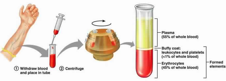
Picture 4 : The image shows the procedures used to collect blood and look for a buffy coat.
Picture source : medical-labs.net
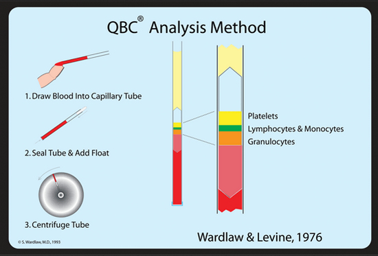
Picture 5 : An analysis of qualitative buffy coat procedure.
Picture source : /i1.wp.com
Buffy coat composition
- White cells
- Platelets
- 10% red blood cells (8, 9)
Buffy coat smear procedure
- Fill the test tube with anti-coagulated blood.
- Centrifuge the blood at about RCF 1000g for about 15 minutes to 25 minutes.
- Remove the supernatant plasma (the fluid above the buffy coat layer).
- Transfer the buffy coat layer and red cells to the slide and mix using a pipette. Make a thin film with the use of a smooth edged spreader.
- Allow a few minutes for the preparation to dry naturally.
- Once the preparation is dry, fix with ethanol for a minute or two.
- Stain using Giemsa staining method or Field’s thin film staining method.
- Examine the preparation under 40X objective and 100X objective.(1, 8, 9, and 10)
References:
- https://en.wikipedia.org/wiki/Buffy_coat
- https://www.stemcell.com/how-to-generate-a-buffy-coat.html
- https://www.sciencedirect.com/topics/immunology-and-microbiology/buffy-coat
- https://microbeonline.com/buffy-coat-definition-composition-preparation-uses/
- https://www.pluriselect.com/knowledge-base/sample-material/buffy-coat.html
- https://www.bioivt.com/biofluids-blood-derived/buffy-coat/
- http://www.bloodjournal.org/content/16/1/1012?sso-checked=true
- https://vcahospitals.com/know-your-pet/buffy-coat-examination-for-mast-cells
- https://jamanetwork.com/journals/jama/article-abstract/197340
- https://content.iospress.com/articles/biorheology/bir25-4-06
Similar Posts:
- Types of Blood Cells
- What is Gram Stain Test – Staining Procedure, Principle and Results
- Difference between T Cells and B Cells
- IMViC Test
- Difference between Mitosis and Meiosis
